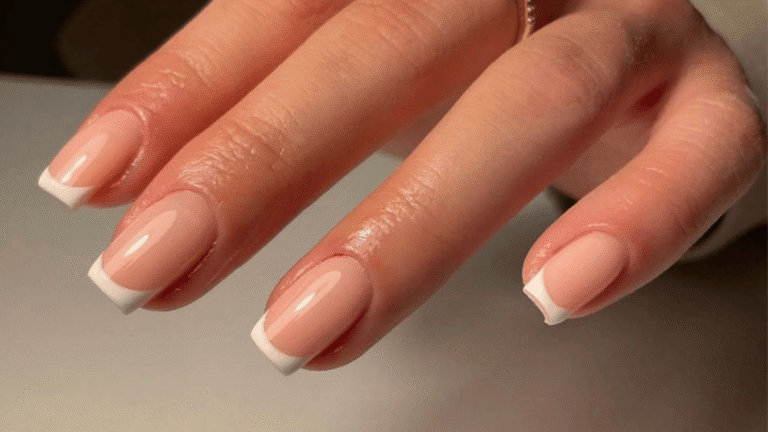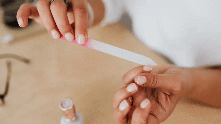

Common yet often overlooked, nail and nail disorders can affect how you function, feel, and present yourself. Changes in nails may signal local issues or a broader disease that needs attention.
This Ultimate Guide outlines causes, symptoms, diagnosis, and treatment for frequent conditions seen in U.S. clinics. You will learn why growth is slow—fingernails take about six months to replace, while toenails may need 12–18 months—and how that timing shapes recovery.
Many problems stem from infections, inflammatory states like psoriasis, or trauma and paronychia. Misdiagnosis is common because different issues can look alike, so accurate evaluation of all 20 nails matters.
Expect evidence-based therapies, from topical and oral medications to simple procedures and home care. Seek care for rapid changes, pain, drainage, or nonhealing lesions to prevent progression and recurrence.
Key Takeaways
- Early evaluation prevents progression and long-term damage.
- Slow nail growth means treatment and visible recovery can take months.
- Similar signs can reflect different conditions, so accurate diagnosis is essential.
- Treatments range from topical care to systemic therapy and procedures.
- See a clinician for pain, drainage, rapid change, or persistent lesions.
What Are Nail Disorders? Understanding Scope and Impact
Changes to the hard plate and surrounding tissues can signal a range of medical problems, from simple infections to systemic disease.
These conditions include problems of the nail plate, bed, and folds that may impair function and quality of life. They are more than cosmetic issues and can make routine tasks—buttoning shirts, typing, or walking—harder.
Clinical findings often overlap across illnesses. That overlap creates frequent misdiagnosis unless clinicians perform a structured assessment, including a detailed history and full 20-nail exam.
Some changes point to local infection or inflammation; others reflect vascular disease, endocrine problems, or even malignancy. The burden rises with age, chronic conditions, and environmental exposures, which increase risk and complicate care.
Early detection, accurate categorization, and targeted therapy improve outcomes. Routine nail checks during medical visits can reveal important clues and reduce long-term complications.
Nail Anatomy 101: Nail Bed, Nail Plate, and Nail Folds Explained
Understanding the unit’s structure clarifies how injuries produce specific signs.
The plate lies over the nail bed and is anchored proximally by the eponychium and laterally by the lateral nail folds. Distally, the hyponychium seals the edge and blocks contaminants.
Proximal versus distal matrix and surface changes
The proximal matrix at the proximal nail bed makes the superficial plate layers. The distal matrix forms the deeper layers.
“Proximal disruption tends to cause superficial pitting; distal injury creates deeper longitudinal ridges.”
That pattern helps clinicians link a visible defect to the matrix location. Superficial pits point to proximal damage, while deep ridging suggests distal matrix involvement.
Folds, cuticle, and protective roles
The cuticle and surrounding fold form a seal that prevents microbes from entering periungual skin. Loss of that barrier raises the risk of paronychia.
- Support: Vascular and connective tissue in the bed give color, attachment, and sensitivity.
- Clinical links: Onycholysis shows lost adhesion to the bed and needs prompt attention.
- Timeline: Fingernails take about six months to replace; toenails need 12–18 months for full growth.
Main Causes of Nail Disorders: Infections, Inflammation, Trauma, and Systemic Disease
Visible plate changes usually reflect one or more root causes. Recognizing patterns helps narrow the differential and point to targeted therapy.
Infections
Onychomycosis is common in older adults and results from dermatophytes, molds, and yeasts. Risk factors include tinea pedis, hyperhidrosis, occlusive footwear, diabetes, and immunosuppression.
Bacterial invasion causes paronychia and Pseudomonas green changes after moisture or trauma breaks the protective seal.
Inflammatory Skin Disease
Psoriasis often produces pitting, onycholysis, and subungual hyperkeratosis. Eczema and lichen planus each have typical plate findings that guide diagnosis.
Trauma and Chemical Exposure
Repeated water exposure, solvents, aggressive manicures, and occupational injury weaken the plate and permit secondary infection.
Underlying Condition Red Flags
Endocrine, vascular, and hematologic disease—such as thyroid dysfunction, peripheral artery disease, and iron deficiency anemia—can cause fragile or slow-growing plates.
- Multiple causes often coexist: trauma can enable infection; inflammation may hide microbes.
- Document exposures and comorbidities to identify the underlying condition and prevent recurrence.
Recognizing Nail Changes and Symptoms
Small shifts in color or texture can signal an early stage of a treatable condition. Early recognition helps guide testing and therapy.
Color, texture, separation, and pain
Patients often present with discoloration, pain, inflammation, or hyperkeratosis. Look for white-yellow or yellow-brown color with subungual debris and thickening — a pattern that often points to fungal involvement.
Common symptom patterns include:
- Discoloration — white, yellow, green, or brown tones.
- Nail pitting and small surface pits.
- Thickening, splitting, and separation from the bed (onycholysis).
- Pain, drainage, or rapid change — consider acute infection or abscess.
Timeline clues: weeks to months
Growth is slow; visible improvement often lags by weeks to months after effective treatment. Asymmetric changes usually suggest a local cause, while symmetric findings raise suspicion for systemic disease.
Tip: Photograph the nails periodically to track progression and response to therapy. Regular images make trends clear and aid clinical decisions.

How Nail Disorders Are Diagnosed Today
Accurate evaluation starts with a focused history and a careful inspection of all twenty plates and surrounding skin.
History and full 20-nail exam
Take a clear timeline: trauma, exposures, systemic diseases, medications, and symptom duration. Examine each plate, the bed, and the cuticle. Check the lateral fold and proximal fold for signs of paronychia or entry points for infection.
Dermoscopy and pattern recognition
Dermoscopy improves bedside accuracy. Look for jagged proximal borders with spikes and longitudinal streaks that suggest fungal invasion versus smooth patterns seen in inflammatory causes.
Laboratory confirmation
Start with in-office KOH (10–20%) for rapid detection. Use PAS histology for higher sensitivity and culture for species identification and viability. PCR offers fast, sensitive organism detection when needed.
When to biopsy
Refer suspicious longitudinal melanonychia—single wide band >3 mm, sudden change, or Hutchinson sign—for dermatology evaluation and biopsy to exclude melanoma.
“Confirm infection before starting systemic therapy to avoid unnecessary risks.”
Nail Disorders
A practical taxonomy helps clinicians sort presentations and prioritize testing. This section groups common problems into infectious, inflammatory, traumatic, and systemic categories to guide initial workup.
- Infectious: onychomycosis, paronychia, and bacterial invasion.
- Inflammatory: psoriasis, lichen planus, and chronic eczematous changes.
- Traumatic: repeated mechanical injury, retronychia, and post‑traumatic dystrophy.
- Systemic: manifestations of endocrine, vascular, or hematologic disease such as Beau’s lines or koilonychia.

Presentations often overlap, so structured differentials and confirmatory testing (KOH, culture, biopsy when indicated) prevent mismanagement. Recognizing hallmark signs speeds referral and treatment.
Upcoming sections will deep dive into high‑prevalence nail conditions with evidence‑based management, timelines, and prevention strategies designed for U.S. clinical practice.
Fungal Nail Infections (Onychomycosis): Causes, Symptoms, and Treatment
Fungal involvement often begins subtly but can progress to widespread thickening and crumbling without targeted therapy. Onychomycosis accounts for roughly half of common plate problems seen in clinics and usually starts in the great toenails.
Risk factors and hallmark signs
Risk rises with older age, tinea pedis, diabetes, immunosuppression, hyperhidrosis, occlusive footwear, and a family history. Typical signs include yellow‑brown discoloration, subungual hyperkeratosis, onycholysis, and distal plate crumbling.
Clinical subtypes
- Distal lateral subungual: most common, starts at the free edge and advances proximally.
- Proximal subungual: rapid proximal spread often seen with weakened immunity.
- White superficial: chalky patches that may scrape off easily.
- Endonyx and total dystrophic: deeper involvement or full destruction of the plate.
Treatment options and testing
Confirm infection before systemic therapy with KOH, PAS, culture, or PCR to guide drug choice. For moderate to severe disease, oral terbinafine is first‑line for dermatophytes; itraconazole or fluconazole are alternatives for yeasts or molds.
Topical agents—efinaconazole, tavaborole, and ciclopirox—are useful for mild cases or when systemic medications are contraindicated, but they need long courses, often 48 weeks or more.
Monitoring, timelines, and prevention
Systemic antifungals require baseline liver testing and review of drug interactions, especially in older adults with comorbidities. Expect slow clearing over months; visible improvement may take a year or longer. Prevent recurrence by treating tinea pedis, keeping footwear dry, and addressing trauma or moisture that breaks the protective seal.
For practical guidance on diagnosis and management, see the Mayo Clinic summary on fungal nail disease — fungal nail treatment and causes.
Paronychia: Acute vs. Chronic Infections of the Nail Fold
Paronychia affects the soft tissues around the plate and can be sudden or long‑standing. Prompt identification guides whether urgent drainage or long‑term control is needed.

Acute presentation and when to drain
Acute paronychia typically follows minor trauma, manicure injury, or biting. It causes rapid erythema, edema, tenderness, and sometimes pus.
Perform incision and drainage if an abscess is present. If no abscess but infection is severe, start oral antibiotics with MRSA coverage in high‑prevalence areas.
Chronic causes and long‑term care
Chronic paronychia lasts more than six weeks and often stems from repeated wet work, irritants, or allergens. Candida superinfection commonly complicates persistent inflammation.
Management centers on strict avoidance of wet work, barrier protection, and restoring the cuticle to protect the nail fold.
Targeted medical therapy
Topical antifungals (eg, ketoconazole) plus low‑potency corticosteroids reduce inflammation and Candida overgrowth. Address Pseudomonas when you see green discoloration; targeted topical therapy such as neomycin may help.
Restore the protective seal of the fold and minimize exposures to prevent recurrence.
Nail Psoriasis: Nail Bed and Nail Plate Changes
Psoriasis often alters both the plate surface and the nail bed beneath, creating distinct, diagnosable signs.
Typical signs and impact
Classic findings include nail pitting and small pitting defects in the plate. Yellow‑brown “oil‑drop” discoloration, subungual hyperkeratosis, separation from the bed, and crumbling or thickening can occur.
These changes can limit function and cause cosmetic concern. Fingernails and toenails may crack, bleed, or snag clothing, reducing hand and foot use.
Treatment options and clinical approach
Differentiate psoriatic dystrophy from fungal infection by exam and targeted testing when unclear. Management blends topical strategies and procedures.
- Topical: potent corticosteroid creams, vitamin D analogs (calcipotriol), and tazarotene for surface pitting and discoloration.
- Procedural: intralesional corticosteroid injections for focal, resistant lesions and laser therapy in selected cases.
Systemic control of psoriasis often improves nail outcomes, but visible recovery takes months due to slow growth.
Brittle Nails (Onychoschizia/Onychorrhexis): Causes from Iron Deficiency to Irritants
Splitting, peeling, and longitudinal ridging commonly cause frustration and limit everyday tasks. These problems often reflect repeated wet work, chemical exposure, or direct trauma to the plate.

Definitions and daily impact
Onychoschizia is lamellar splitting — thin layers peel from the free edge. Onychorrhexis means vertical fissures that weaken the plate and snag clothing.
Idiopathic vs secondary causes
Some people have an idiopathic fragility without clear triggers. Others have a secondary condition such as psoriasis, eczema, lichen planus, vitamin deficits, or iron deficiency.
Certain medications, including retinoids, some antiretrovirals, and chemotherapeutics, may worsen brittleness.
Practical care and prevention
- Minimize water and solvent exposure; use cotton under vinyl gloves for wet tasks.
- Keep plates short and trim to reduce leverage and splitting.
- Apply emollients and humectants (glycerin or propylene glycol) daily to hydrate the plate and surrounding skin.
- Use nail hardeners cautiously; screen for iron and other nutritional gaps when fragility is severe.
Biotin is commonly used but has limited evidence and can interfere with lab tests; discuss use with your clinician.
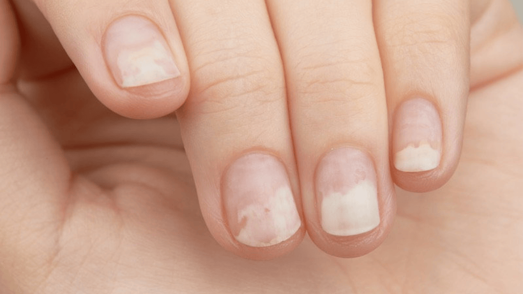
Onycholysis: When the Nail Separates from the Nail Bed
Onycholysis presents as a painless lifting of the plate that typically develops slowly. Patients often note a pale trailing edge and debris that darkens from trapped material or secondary colonization.
Common triggers and causes
Mechanical and chemical factors include minor trauma, excessive filing, prolonged water exposure, and contact with solvents or irritants.
Systemic causes include psoriasis, fungal infection, and reactions to certain medications.
Evaluation
Assess recent injury, workplace exposures, and new drugs. Inspect for discoloration or thickening that suggests coexisting onychomycosis.
Confirm infection with KOH, culture, or PAS when results will change management.
Management and home care
Treat the underlying cause: manage psoriasis, correct deficiencies, or give oral antifungals for confirmed fungal disease. Protect the plate by trimming the detached edge and avoiding aggressive filing.
- Keep the area dry and avoid prolonged wet work; wear gloves for exposure.
- Avoid harsh chemicals and separate tools for affected plates.
- Follow clinician advice for specific medical treatment and expect slow reattachment over months.
“Eliminating triggers and protecting the plate are key steps to support regrowth and reattachment.”
Ingrown Toenails: Prevention, Symptoms, and Treatment Options
Ingrown toenails begin when the plate edge pierces adjacent soft tissue, triggering pain and inflammation. Genetics, tight socks or shoes, improper trimming, and direct trauma all raise risk.
Early signs and when to seek care
Look for swelling, tenderness, redness, soreness, or pus near the lateral edge. Mild cases respond to home care, but severe pain, spreading redness, or drainage needs prompt clinical review for possible infection or cellulitis.
Home care vs surgical approaches
Start conservative care: warm water soaks 3–4 times daily, comfortable footwear, keep feet dry, and oral analgesics as needed.
- Prevention: trim straight across, avoid rounding edges, and choose roomy shoes.
- Conservative: packing with cotton, topical antiseptics, and temporary elevation of the lateral tissue.
- Surgical options: partial avulsion or matrix procedures remove the offending edge for recurrent or severe cases.
“Escalate care when pain or drainage suggests deep infection; antibiotics are considered if cellulitis or purulent drainage is present.”
For practical clinical guidance, review the Mayo Clinic guidance and everyday foot care tips.
Thickened and Overgrown Nails (Onychogryphosis)
Severe overgrowth of the big toe plate often creates a curved, ram’s‑horn deformity that impairs walking and shoe fit. This condition produces marked thickening and can trap debris, irritate surrounding tissue, and raise infection risk.
Contributors and common causes
Genetic tendency, repeated pressure or trauma, poor circulation, psoriasis, and ichthyosis all contribute. Chronic neglect or inadequate footwear accelerates deformity and functional loss.
Care pathways and practical steps
Conservative care focuses on periodic professional debridement and reduction by a podiatrist to restore comfort and prevent soft‑tissue injury. Regular trimming and cushioning improve mobility and reduce pain.
Definitive options when conservative care fails
For severe deformity or recurrent problems, surgical nail bed ablation or partial avulsion offers a permanent solution. Discuss risks and recovery with a foot specialist before proceeding.
“Periodic professional trimming and timely surgical referral prevent ulceration and restore function.”
- Protective measures: roomy shoes, orthotic padding, and moisture control.
- Monitor: check the surrounding bed and tissue for redness or drainage.
- When to refer: persistent pain, recurrent infection, or inability to ambulate comfortably.
Nail Signs That May Indicate Systemic Disease
Certain plate findings can be clinical red flags for systemic disease. Recognizing them prompts timely workups and targeted therapy.

Beau’s lines, clubbing, koilonychia, and splinter hemorrhages
Beau’s lines are transverse grooves that reflect interruption of mitosis in the nail bed after severe illness or high fever. Their distance from the proximal edge helps time the event.
Clubbing involves soft-tissue enlargement at the digit with an increased Lovibond angle and often signals cardiopulmonary or other systemic diseases.
Koilonychia (spooning) links to iron deficiency and merits evaluation for anemia and its sources.
Splinter hemorrhages are thin, linear bleeding under the plate and can follow trauma; multiple or proximal lesions raise concern for endocarditis or vasculitic conditions.
Longitudinal melanonychia and pincer deformity: when to refer
Longitudinal melanonychia may be benign, but a single wide band, rapid change, or Hutchinson sign requires dermatology referral and possible biopsy to exclude subungual melanoma.
Pincer nail—excessive transverse curvature—associates with psoriasis, onychomycosis, SLE, and some drugs. Consider surgical correction for severe pain or recurrent soft‑tissue injury.
When plate signs coexist with systemic symptoms or risk factors, broaden the evaluation to uncover underlying disease.
Treatment Toolbox: Medications, Procedures, and At-Home Care
Combining evidence‑based medications, selected procedures, and practical self‑care gives the best chance of recovery. This toolbox helps clinicians and patients weigh options, balance risks, and plan follow‑up.
Medications: classes and monitoring
Antifungals — oral terbinafine is favored for dermatophyte infection, especially in older adults; itraconazole works better for yeasts or molds, and fluconazole is an alternative. Topical agents (efinaconazole, tavaborole, ciclopirox) require long courses.
Corticosteroids and antibiotics treat inflammatory causes and acute bacterial flares. Monitor for drug interactions and hepatic safety when using systemic agents.
Procedures: targeted interventions
Incision and drainage manages abscessed paronychia and relieves pain quickly. Partial or total avulsion helps severe dystrophy or recurring problems. Laser therapy may be an adjunct for resistant fungal disease or select psoriatic changes.
At‑home measures and relapse prevention
Control moisture, avoid irritant exposure, and protect folds with barrier creams. Use cotton under vinyl gloves for wet work and practice safe trimming and salon hygiene.
“Match the chosen treatment to the cause, the comorbidity profile, and the patient’s goals.”
Think acute versus chronic—address active infections promptly, and plan long‑term strategies for relapsing inflammatory conditions and recurrent infections.
Prevention and Nail Care Best Practices for Healthy Nails
Small routine changes protect the plates and surrounding skin from common harms. Prevention focuses on reducing moisture, limiting trauma, and keeping tools and footwear clean.
Glove use, trimming techniques, and salon hygiene
Glove protocol: Use cotton liners under vinyl or nitrile gloves for wet tasks to cut repeated exposure and friction. Replace liners when wet.
Trimming: Trim toenails straight across and avoid rounded edges to prevent ingrown edges. File gently to smooth rough tips and reduce splitting.
Salon safety: Choose salons that sterilize instruments, avoid aggressive cuticle removal, and refuse services if tools look unclean.
Managing moisture, hyperhidrosis, and footwear risk
Keep feet dry by using moisture-wicking socks and breathable shoes. Change socks within a day if they become damp.
For excessive sweating, consider topical antiperspirants, foot powders, or a clinician review for hyperhidrosis treatments.
- Inspect plates every few weeks to catch early changes and act quickly.
- Barrier creams and emollients protect the folds during repetitive chemical or water use.
- Shoe choices: Pick roomy, ventilated footwear to reduce fungal growth risk.
“Prevention is faster and simpler than treatment—small steps reduce recurrence and support long-term health.”
When to See a Dermatologist or Podiatrist in the United States
Seek specialist care when changes occur quickly, are painful, or fail to heal with routine measures.
Warning signs that need prompt evaluation
Urgent features include rapid color change, new severe pain, visible pus or drainage, and green discoloration that suggests Pseudomonas. Also seek care for nonhealing lesions or sudden widening of a pigmented band.
Suspected abscess in paronychia requires procedural drainage by a clinician. Persistent or worsening infections despite treatment merit specialist assessment and possible culture or imaging.
Special populations who need earlier referral
People with diabetes, peripheral vascular disease, or immunosuppression face higher risk for complications. In these cases, refer sooner rather than later to avoid cellulitis, ulceration, or deep infection.
When advanced diagnostics or biopsy are needed
Recurrent infections, treatment failures, or atypical presentations prompt advanced testing (KOH, culture, PCR, PAS) or biopsy. Concerning longitudinal pigment—especially a single wide band or rapid change—requires dermatology biopsy to exclude melanoma.
- Urgent specialist features: rapid change, pain, drainage, green color, abscess.
- High-risk groups: diabetes, immunosuppression, poor circulation—refer early.
- Triggers for advanced workup: recurrent cases, treatment failure, atypical signs.
- Follow-up timing: expect serial checks over weeks to months because regrowth is slow.
Use primary care for initial triage and ask for dermatology or podiatry referral when urgent signs or high-risk features are present. For routine foot care guidance, review practical tips at foot care tips.
“Early specialist input for red flags reduces morbidity and shortens recovery time.”
Conclusion
Timely evaluation and targeted care make the biggest difference when visible changes appear on fingers or toes. Nail disorders are common and can reflect local injury or a broader disease. Careful assessment helps link signs to underlying causes and plan effective treatment.
Accurate diagnosis avoids unnecessary therapies and reduces risk. Management combines topical and systemic treatment, procedures, and clear self-care steps.
Prevention and adherence matter because visible recovery mirrors slow plate development. Keep routine checks, protect the plate from trauma, and address moisture and exposures early.
Collaborate with clinicians for persistent or complex conditions. Prompt escalation for red flags preserves function, limits harm, and supports long-term quality of life.
FAQ
What causes changes in the nail bed, plate, or folds?
How do I tell acute paronychia from chronic paronychia?
When should I worry about nail pitting or longitudinal lines?
What tests confirm a fungal infection of the nails?
Are oral antifungals safe and how long do they take to work?
How is chronic paronychia treated long term?
What causes brittle, splitting nails and how can I improve them?
When is nail avulsion or surgical care necessary?
Can medications or systemic disease cause onycholysis or thickening?
How do I prevent recurrent fungal or bacterial infections of the toes and folds?
What are red flags that require urgent dermatology or podiatry referral?
Can topical treatments alone clear fungal toenail infections?
How does psoriasis affect the nail plate and surrounding tissue?
What self-care helps healing after incision and drainage of an abscess?